top of page

Corner Tumors and Trigeminal Neuralgia

What is a cerebellum? A cerebellum is a specific term referring to the lateral side of the posterior fossa in the brain. The petrous bone, a strong pyramid-shaped bone containing the structures of the inner ear, forms an approximately ninety-degree angle with the cerebellum, a thick membrane called the tentorium (meaning tent in English), which separates the cerebellum from the base of the brain. In this area, located on the lateral edge of the cerebellum, lies the groove between the pons, the egg-shaped, rounded part of the brainstem, and the cerebellum.
This area, where the cerebellum and pons intersect and the petrous bone and tentorium form a corner, contains the 5th cerebral nerve, the 7th and 8th cerebral nerve complexes, and the 9th, 10th, and 11th cerebral nerve complexes. Two important vessels supplying the cerebellum, the AICA and PICA, are also located in this area. The 6th cerebral nerve is also found deep within this area. Thus, when we refer to this corner, we are talking about an extremely complex anatomical region. Tumors, cystic lesions, and various vascular and nerve diseases can occur in this corner region.

The most common lesions located in the corner are:

Corner tumor: vestibular schwannoma 80-90%

Corner meningioma 5-10%

Corner ependymoma

Corner epidermoid cyst

Tumors of the petrous apex

Angular arachnoid cyst

Trigeminal neuralgia

Hemifacial spasm

Glossopharyngeal neuralgia

We use neuromonitoring in facial nerve angle tumor surgeries . We do this to prevent facial paralysis. During the surgery, the neuromonitoring team monitors the facial expressions made by the facial nerve, both audibly and on the screen. However, although neuromonitoring is very useful, it is not always a system that prevents facial paralysis. Despite all precautions, facial paralysis can sometimes develop in facial nerve angle tumors. If facial paralysis develops after facial nerve angle tumor surgery, we wait 3 months, and if there is no improvement, we perform facial resuscitation surgery.

CPA_angle_tumor_prof_dr_bulent duz

Köşe Tümörü Ameliyatı

Corner Tumor: My male patient, pictured above, had a corner tumor located on the right side. It was quite large, filling the entire corner and compressing half of the brainstem with its cyst. I surgically removed the tumor and its cyst. The pathology report confirmed schwannoma. The patient experienced no neurological regression after surgery. There was no facial paralysis. In fact, his hearing was preserved; he can hear conversations from close range after the surgery, but he cannot understand distant sounds.

PCA_tumor_right_Prof_Bulent_Duz
PCA_tumor_right_after_operation

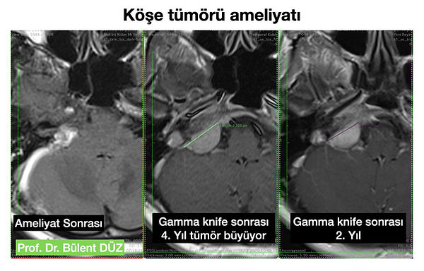
A 42-year-old female patient came to my practice having undergone radiotherapy four years prior for a cranial nerve tumor. She had initially presented with tinnitus. Despite undergoing Gamma Knife treatment over the past four years, the tumor had grown, and she was experiencing balance problems, impaired arm coordination, dizziness, and worsening tinnitus. A cranial nerve tumor becomes more adherent to surrounding tissue after Gamma Knife treatment, making surgery more difficult. Removing a tumor so extensively embedded in the petrous bone at the base of the skull is quite challenging. The greatest risk is facial paralysis. I operated on my patient and removed the tumor almost completely microscopically. The tumor was severely adherent to the facial nerve. I separated the tumor from the nerve and, under neuromonitoring control, removed it without damaging the nerve. After the surgery, my patient was smiling and had no facial paralysis; she left the operating room without experiencing any facial paralysis.

What are the symptoms of corner tumor disease? What are the risks of corner tumor surgery? You can watch the video above.

Familiarity with the anatomy of the pons is essential for performing surgery on pons tumors. All pons anatomy includes the facial and vestibulocochlear nerves. The trigeminal nerve is located above the 7th-8th complex, while the hypoglossal nerve, vagus nerve, and accessory nerve are located below it. Understanding the specific placement and appearance of these nerve complexes is crucial for pons surgery. Further up is the trochlear nerve, which is the most critical structure in the pons anatomy. Although not vascularly visible here, the pica above and aica below are also encountered in many surgeries. The most common pons tumors are acoustic schwannomas, meningiomas, and epidermoid tumors. Acoustic schwannomas mostly originate from the inferior vestibular nerve. In acoustic schwannoma surgery, knowing the spatial position of the facial nerve relative to the tumor is necessary to minimize facial nerve damage and prevent facial paralysis. This is best understood with diffusion tensor imaging of the facial nerve combined with MR tractography.

Angular Meningioma: Above, I showed a case of a meningioma located in the cerebellopontine angle. Angular meningiomas are usually only noticed after they have grown, because they do not cause any symptoms while growing. By the time they are noticed, they are pressing on the facial nerve, auditory nerve, and brainstem. Above, I have prepared an instructional video showing how I performed surgery on a 3 cm meningioma originating from the tentorium, pushing against the neurovascular structures in the angle and adhering to the AICA. I hope those interested in microsurgery will enjoy watching it.

Corner Meningioma: Above, I showed a case of a meningioma located in the petrous apex. Tumors that originate from the anterior side of the trigeminal nerve and grow by pushing the trigeminal nerve backward are called petrous apex tumors. Here, I showed how I performed surgery on a patient with a giant meningioma that originated from the corner in the posterior fossa, then progressed to the central fossa, and from there entered the maxillary sinus in the face.

I have shown the anatomy of the cranial angle in detail endoscopically in my presentation above. I went into the surgery above with a preliminary diagnosis of arachnoid cyst, but ultimately encountered a Rathke's Cleft cyst in the cranial angle, a very rare condition. In this case, the anatomy of the cranial angle and the relationship between the cranial nerves located in the cranial angle are shown in a highly instructive way.

Trigeminal neuralgia - The maddening disease

Trigeminal neuralgia is a disease that causes excruciating pain, usually resulting from compression of the trigeminal nerve (the 5th cranial nerve) by a blood vessel called the AICA. Trigeminal neuralgia surgeries can be performed both microscopically and endoscopically. I have shown examples of how I perform endoscopic trigeminal neuralgia surgeries below. Learning about trigeminal nerve anatomy is an excellent resource for understanding trigeminal nerve anatomy.

RF Rhizotomy Procedure for Trigeminal Neuralgia: I performed radiofrequency rhizotomy on my 68-year-old patient with an epidermoid tumor after surgery. The video below shows how I performed the corner tumor surgery using microscopic and endoscopic approaches, followed by the RF rhizotomy procedure.

Prof. Dr. Bülent DÜZ

All rights reserved. No part of this content may be copied, in whole or in part, without permission. The content is for informational purposes only and may not be used for other purposes.
Reproduction, electronic or otherwise, without the permission of Bulent Düz MD is strictly forbidden.

All rights reserved 2026

bottom of page